Case Reports
Case 4 : Schistosomiasis of Urinary Bladder
Contributed by :Dr Kalpesh Khatal,and Dr Anirudh Badade
A 29 year old female was presented with increased frequency of urination and tiny particles passing through urine.
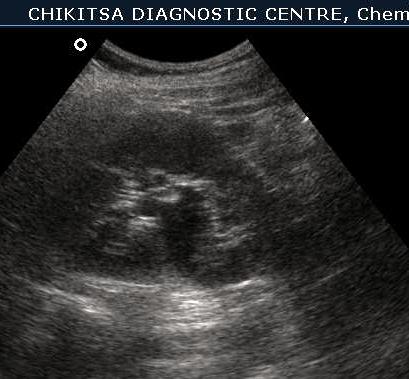
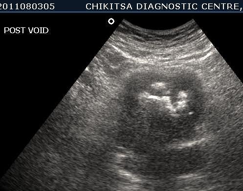
Ultrasound Images:
March 2012 :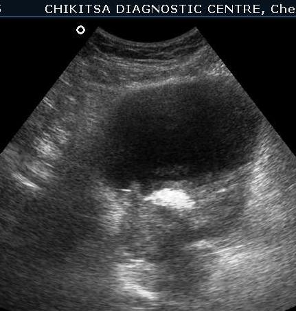
5 5 cm x 5 cm size ill-defined heterogeneous area in the posterior wall of the urinary bladder which is thickened and measures upto 0.6 cm in thickness. Large solid calcific areas are noted in the thicknened portion of the wall urinary bladder. Suggestive of schistosomiasis of the urinary bladder (possibly with malignant change).
Left kidney shows hydronephrosis.
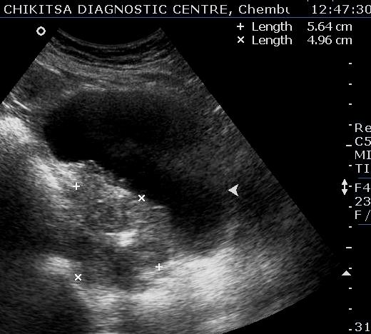
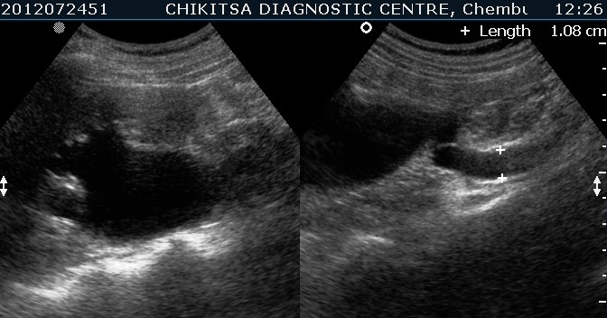
July-Aug 2012 :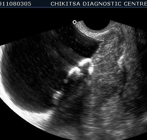
Left kidney shows hydronephrosis.
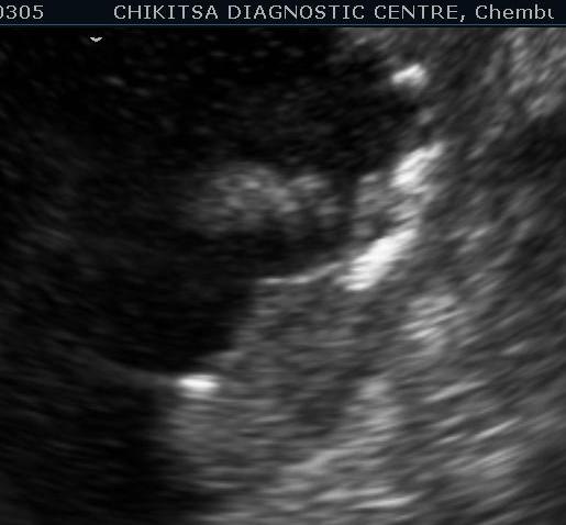
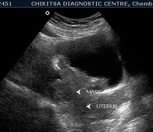
Lesion in the wall of the urinary bladder has increased in size. Calcific areas are noted. Urinary bladder malignancy ( confirmed by histopathology), possibly following schistosomiasis.
Lesion in the wall of the urinary bladder has increased in size. Calcific areas are noted. Urinary bladder malignancy ( confirmed by histopathology), possibly following schistosomiasis.
DISCUSSION :-
Genitourinary schistosomiasis is produced by Schistosoma haematobium, a species of fluke that is endemic to Africa and the Middle East, and causes substantial morbidity and mortality in those regions. It also may be seen elsewhere, as a result of travel or immigration.
Schistosoma haematobium, one of the five fluke species responsible for most human schistosomiasis, is the only species that infects the genitourinary system.
In the early stages, it involves the bladder and ureters; later, the kidneys and genital organs .
The larvae of Schistosoma haematobium are released from snails into water and penetrate human skin exposed to the infected water. These larvae travel to the lungs and liver where they reside and mature. After maturation, pairs of adult worm travel to the pelvic veins. Eggs are deposited in the bladder wall vessels and cause a granulomatous response resulting in polypoid lesions.
Ultrasound featrures : mucosal irregularity, polypoidal bladder wall thickening, inflammatory pseudopolyps (with increased vascularity on Doppler), ureteral and pelvicalyceal system dilatation (mainly if ureterovesical junction involved), stricture, curvilinear dense wall calcification. Later, bladder capacity may be reduced; rarely ureteritis cystica.
The calcification in bladder wall is due to calcification of the ova deposited in the bladder wall vessels. The calcification spreads around the bladder wall and may completely encircle the bladder, appearing as a curvilinear ring. The bladder wall becomes fibrotic and later bladder capacity may be reduced.
A bladder carcinoma may be considered when follow-up imaging shows an absence of wall calcification in areas that were previously calcified.
Heavy egg deposits in the bladder mucosa and submucosa act as a mechanical irritant to the urothelium, inducing chronic inflammatory lesions, which causes hyperplasia and squamous metaplasia and eventually carcinoma.
Many mechanisms have been proposed to explain the malignant change.
Acknowledgment:- I am thankful to Dr Anirudh Badade for the support and encouragement whenever I was in need.
References
1) Radiographics. 2012 Jul-Aug;32(4):1031-46.
2) Kuper H, Adami HO, Trichopoulos D. Infections as a major preventable cause of human cancer. J Intern Med. 2000;248:171-183.
3) Cheever A W. Schistosomiasis and neoplasia. J Natl Cancer Inst. 1978;61:13-18.
4) Cheever A W, Kuntz R E, Moore J A, Hang T C. Pathology of Schistosoma haematobium infection in the Capuchin monkey. Trans R Soc Trop Med Hyg. 1988;82:107-111.
5) Christie J D, Crous D, Kelada A S, Anis-Ishak E, Smith J H, Kamel I A. Patterns of Schistosoma haematobium egg distribution in the human urinary tract. III. Cancerous lower urinary tracts. Am Trop Med Hyg. 1986;35:759-764
6) Hicks R M, James C, Webbe G. Effect of Schistosomiasis haematobium and N-butyl-N-(4-hydroxybutyl)nitrosamine on the development of urothelial neoplasia in baboon. Br J Cancer. 1980;42:730-755
7) Ishikawa J, Xu H J, Hu X S, Yandell D W, Maeda S, Kamidono S, Benedict W F, Takahashi R. Inactivation of the retinoblastoma gene in human bladder and renal-cell carcinomas. Cancer Res. 1991;51:5736-5743
8) Knowles M A, Williamson M. Mutation of H-ras is infrequent in bladder cancer: confirmation by single-strand-conformation-polymorphism analysis, designed restriction-fragment-length polymorphisms, and direct sequencing. Cancer Res. 1993;53:133-139.
9) Sidransky D, Von Eschenbach A, Tsai Y C, Jones P, Summerhayes I, Marshall F, Pual M, Green P, Vogelstein B. Identification of the p53 gene mutations in bladder cancers and urine samples. Science. 1991;252:706-709